As an expert in anatomy and physiology, I’ve often been asked about the joints that are most affected in the human body. Today, I’ll be diving into this topic and exploring which specific structure in the figure is most prone to joint issues. Understanding the vulnerabilities of our joints is crucial for maintaining overall health and preventing potential complications. So, let’s take a closer look at the figure and uncover the joints that require extra care and attention.
When it comes to joint health, knowledge is power. That’s why I’m excited to discuss the structure in the figure that is particularly susceptible to joint problems. By identifying this area, we can better understand the importance of proper care and potentially prevent future issues. So, stay tuned as I reveal the key joint that you should be mindful of and share some valuable insights on how to keep it in optimal condition.
We all know that joints play a vital role in our everyday movements, but have you ever wondered which specific structure in the figure is most prone to joint-related complications? Today, I’ll be shedding light on this intriguing topic and providing you with the essential information you need to know. By understanding the joints that are most affected, we can take proactive steps to maintain their health and functionality. So, let’s explore the figure and discover the key structure that demands our attention.
Which Structure in the Figure is the Primary Area of Degeneration in Osteoarthritis?
When it comes to maintaining overall health and preventing complications, understanding the structure of our joints is crucial. Joints are the connections between our bones that allow for movement and flexibility. They play a vital role in our day-to-day activities, whether it’s walking, running, or even simple tasks like reaching and grabbing objects.
The human body has numerous joints, each with its own unique structure and function. These joints can vary in complexity, ranging from simple hinge joints like the knee and elbow, to more complex ball-and-socket joints like the hip and shoulder. Regardless of their complexity, all joints are susceptible to wear and tear over time, making it important to take care of them to maintain optimal function.
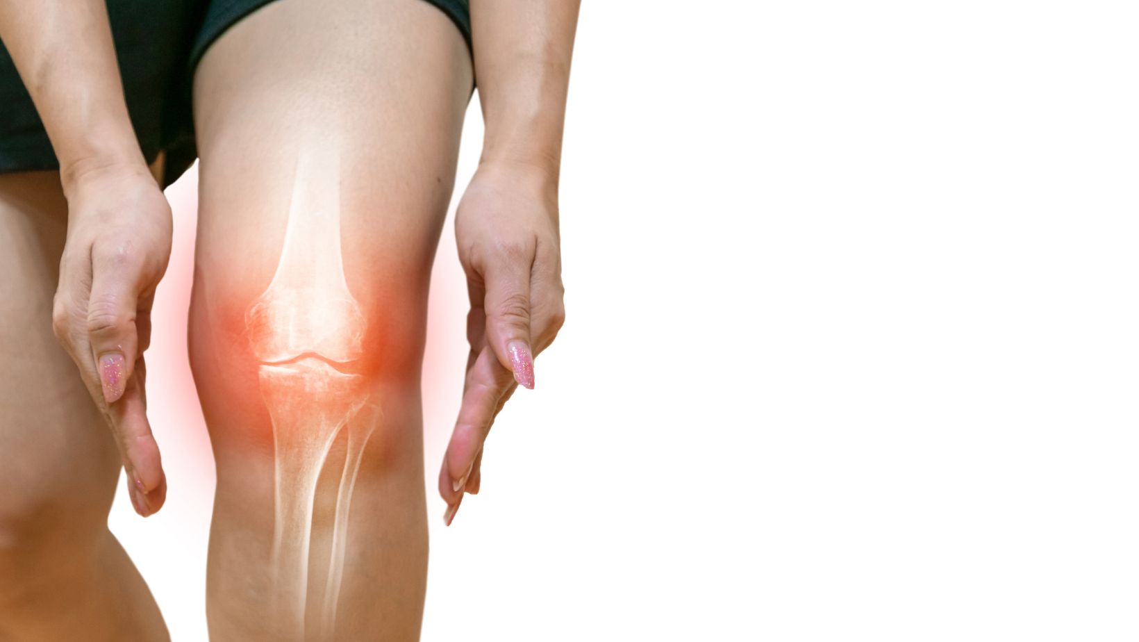
One joint structure in particular is prone to more issues than others: the knee joint. The knee joint is a hinge joint that connects the thighbone (femur) to the shinbone (tibia). It is responsible for bearing a significant amount of our body weight and plays a crucial role in walking, running, and other weight-bearing activities.

The Anatomy of Joints
Understanding the structure of our joints is essential for maintaining overall health and preventing complications. Joints are where two or more bones come together, allowing us to move our bodies in various ways. The human body has many different types of joints, each with its own unique structure and function.
- Synovial Joints: These are the most common type of joints in the body and are responsible for our ability to move freely. Examples of synovial joints include the knee, elbow, and shoulder joints. They are characterized by the presence of a joint cavity filled with synovial fluid, which helps reduce friction and allows for smooth movement.
- Cartilaginous Joints: These joints are found where bones are connected by cartilage. They provide more stability than synovial joints but have limited movement. Examples of cartilaginous joints include the joints between the vertebrae in the spine and the pubic symphysis.
- Fibrous Joints: These joints are held together by strong connective tissue, such as ligaments or fibrous capsules. They provide little to no movement and primarily serve to hold bones together. Examples of fibrous joints include the sutures in the skull and the joint between the tibia and fibula in the lower leg.
Conclusion
Understanding the most commonly affected joints and the structures within them that are prone to injury is crucial for maintaining joint health and preventing injuries. In this article, I have highlighted the knees, ankles, shoulders, and wrists as the joints that are most frequently affected. By being aware of the various injuries that can occur in these joints and recognizing the associated symptoms, individuals can take proactive measures to protect themselves.
To prevent joint injuries, it is important to maintain proper form during physical activities, wear protective gear when necessary, and avoid overuse or repetitive motions. Engaging in regular exercise that includes strengthening and stretching exercises is also essential for keeping joints strong and flexible. By following these recommendations, individuals can reduce the risk of joint injuries and enjoy an active lifestyle.


